

Pterygia (singular pterygium) and pinguecula (singular pinguecula) are benign tissue growths that can develop on the eye's surface and, in some cases, on the cornea (the transparent dome-shaped layer that covers the eye). Fairly common growths, pterygia, and pinguecula primarily affect adults who spend a significant amount of time outdoors and in the sun. However, pterygia and pinguecula1 can appear in anybody, including children.
Although these two types of noncancerous growths have many symptoms in common, they have subtle differences, mainly related to their growth pattern. Whereas a pinguecula2 occurs only on the conjunctiva (the protective membrane that covers the white of the eye), a pterygium3 can also grow across the cornea. At Beach Eye Medical Group, our Orange County eye doctors have vast experience in the diagnosis and management of both conditions.

Pterygia and pinguecula are generally associated with environmental factors such as ultraviolet (UV) light, wind, a dry climate, and dust; however, the precise cause of these eye growths has not been identified.
For a proper diagnosis, the eye should be evaluated by one of our board certified eye doctors. To examine the eye, our Beach Eye Medical Group specialists will use a device called a slit-lamp to detect abnormalities. A slit lamp emits a narrow beam of light and provides a magnified three-dimensional view of the eye, which allows us to confirm the presence of a pterygium or pinguecula. Once a diagnosis is confirmed, treatment can be recommended.
When asymptomatic, the growths generally do not require any treatment. If a pterygium or pinguecula becomes inflamed or irritated, eye drops or ointments may be prescribed to alleviate discomfort. If the growths become large enough to encroach on the cornea and threaten vision, removal will be necessary. Some people may also opt for removal for cosmetic reasons.
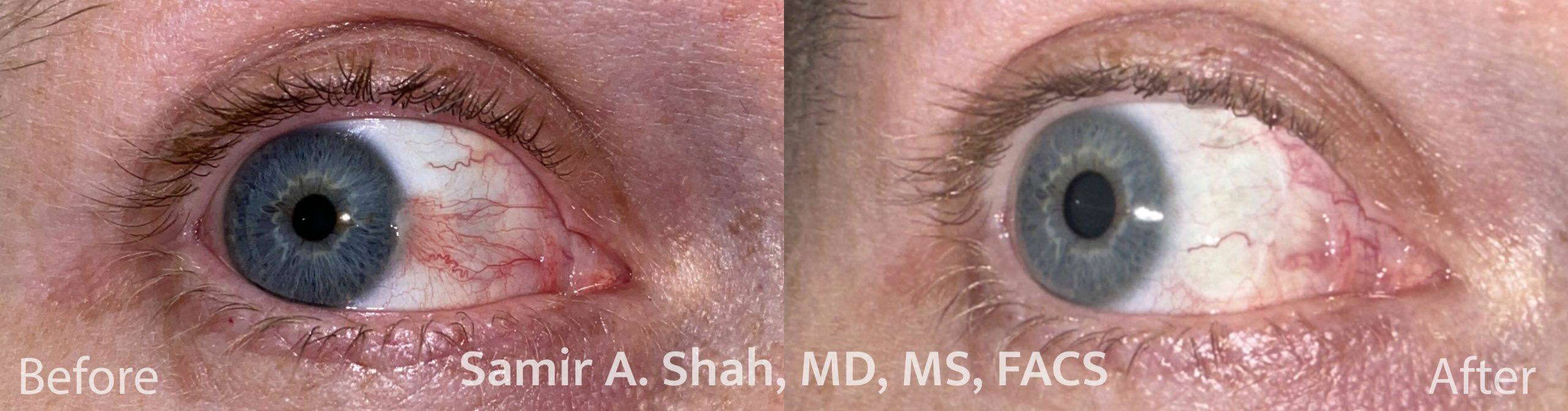
If surgery is needed or desired, our Orange County eye surgeons can remove the growth in a standard outpatient procedure. During the procedure, a topical anesthetic is applied to the eye's surface and the growth is excised. The surgery is painless and the patient's eyes are open during the process. Typically, the procedure takes about half an hour to complete. Although surgery can be highly effective, recurrence happens in about a third of patients seeking removal. To help prevent regrowth, our eye surgeons use a suturing or adhesive technique to graft tissue onto the treatment area. A course of growth-discouraging steroid drops may also be recommended as part of the post-surgical care.
Patients are advised to take a day off on surgery date. Downtime is minimal and patients can expect to return to their normal routines a day after the procedure. To protect the eye, it is recommended that the patient wears an eyepatch for up to two days following the surgery. During the recovery period, patients generally use special drops to promote the healing process and reduce any inflammation associated with the procedure.
To decrease the possibility of recurrence, patients should routinely and adequately protect the eyes against UV light and dust, as well keep the eyes well lubricated when in dry environments. Wraparound sunglasses can be worn for protection and artificial tears can be used to add moisture to the eyes.
There are very few risks associated with removal; however, there is a small chance of astigmatism, particularly in those who already have this optical condition. All pre-existing eye conditions should be discussed with our doctors before surgery.
Click here to watch Dr. Shah perform a pterygium excision.
References
1 What Is a Pinguecula and a Pterygium (Surfer's Eye)? Available: https://www.aao.org/eye-health/diseases/pinguecula-pterygium
2 Pinguecula. Available: https://www.ncbi.nlm.nih.gov/books/NBK558965/
3 Understanding and managing pterygium. Available: https://www.ncbi.nlm.nih.gov/pmc/articles/PMC5340105/
4 What Is Surfer's Eye? Available: https://www.webmd.com/eye-health/pterygium-surfers-eye
Always a first class, profession visit with Beach Eye Medical Group. They always treat me the way we all hope to be treated.
Medically Reviewed by
Samir Shah, MD
Samir Shah, MD, MS, FACS, is a board-certified ophthalmologist specializing in custom laser cataract surgery and refractive lens exchange. A Johns Hopkins fellow and UCLA alumnus, he is nationally recognized as an expert in refractive cataract surgery. Recently, he was named the Top Doctor 2026 by the Orange County Medical Association and Orange Coast Magazine.