
The retina is the light-sensitive tissue lining the back of our eye. Light rays are focused onto the retina through our cornea, pupil and lens. The retina converts the light rays into impulses that travel through the optic nerve to our brain, where they are interpreted as the images we see.
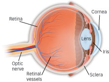
The middle of our eye is filled with a clear gel called vitreous (vit-ree-us) that is attached to the retina.
Sometimes tiny clumps of gel or cells inside the vitreous will cast shadows on the retina, and you may sometimes see small dots, specks, strings or clouds moving in your field of vision. These are called floaters. You can often see them when looking at a plain, light background, like a blank wall or blue sky.
As we get older, the vitreous may shrink and pull on the retina. When this happens, you may notice what look like flashing lights, lightning streaks or the sensation of seeing “stars.” These are called flashes.
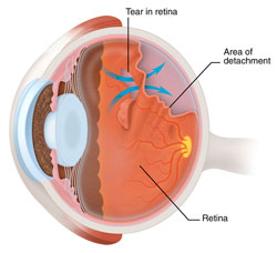
Usually the vitreous moves away from the retina without causing problems. But sometimes the vitreous pulls hard enough to tear the retina in one or more places. Fluid may pass through a retinal tear, lifting the retina off the back of the eye — much as wallpaper can peel off a wall. When the retina is pulled away from the back of the eye like this, it is called a retinal detachment1.
The retina does not work when it is detached and vision is blurry. A retinal detachment2 is a very serious problem that almost always causes blindness unless it is treated.
Symptoms of both retinal tears and retinal detachments can include the following:
Floaters and flashes in themselves are quite common and do not always mean you have a retinal tear or detachment. However, if they are suddenly more severe and you notice you are losing vision, you should book an urgent appointment with at our Beach Eye Irvine office right away.
People with the following conditions have an increased risk for retinal detachment:
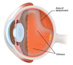
The middle of the eye is filled with a clear gel called vitreous (vit-ree-us) that is attached to the retina. As we get older, the vitreous may shrink and pull away from the retina.
Usually the vitreous separates from the retina without causing problems. But sometimes the vitreous pulls hard enough to tear the retina in one or more places. Fluid may pass through the retinal tear, lifting the retina off the back of the eye, much as wallpaper can peel off a wall.
At Beach Eye Medical Group, our board certified ophthalmologists can diagnose retinal tear or detachment during an eye examination where he or she dilates (widens) the pupils of your eyes. Some retinal detachments are found during a routine eye examination.
Only after careful examination can your ophthalmologist tell whether a retinal tear or early retinal detachment is present.
Most retinal tears need to be treated by sealing the retina to the back wall of the eye with laser surgery or cryotherapy (a freezing treatment). Both of these procedures create a scar that helps seal the retina to the back of the eye. This prevents fluid from traveling through the tear and under the retina, which usually prevents the retina from detaching. These treatments cause little or no discomfort and may be performed in our Irvine office.
Almost all patients with retinal detachments must have surgery to place the retina back in its proper position. Otherwise, the retina will lose the ability to function, possibly permanently, and blindness can result. The method for fixing retinal detachment depends on the characteristics of the detachment. In each of the following methods, our ophthalmologists. will locate the retinal tears and use laser surgery or cryotherapy to seal the tear.
This treatment involves placing a flexible band (scleral buckle) around the eye to counteract the force pulling the retina out of place.
The ophthalmologist often drains the fluid under the detached retina, allowing the retina to settle back into its normal position against the back wall of the eye. This procedure is performed in an operating room.
In this procedure, a gas bubble is injected into the vitreous space inside the eye in combination with laser surgery or cryotherapy. The gas bubble pushes the retinal tear into place against the back wall of the eye. Depending on the severity, this procedure can be completed at the Beach Eye Irvine office.
Your ophthalmologist will ask you to constantly maintain a certain head position for several days. The gas bubble will gradually disappear.
This surgery is performed in an operating room. The vitreous gel, which is pulling on the retina, is removed from the eye and usually replaced with a gas bubble. Your body's own fluids will gradually replace the gas bubble. Sometimes vitrectomy is combined with a scleral buckle.
Most retinal detachment surgery is successful, although a second operation is sometimes needed.
Some retinal detachments cannot be fixed. If the retina cannot be reattached, the eye will continue to lose sight and ultimately become blind.
After successful surgery for retinal detachment, vision may take many months to improve and, in some cases, may never return fully. Unfortunately, some patients do not recover any vision. The more severe the detachment, the less vision may return. For this reason, it is very important to see your ophthalmologist at the first sign of any trouble.
References
1 Detached Retina. Available: https://www.aao.org/eye-health/diseases/detached-torn-retina
2 Corneal Conditions. Available: https://www.nei.nih.gov/learn-about-eye-health/eye-conditions-and-diseases/corneal-conditions
3 Nearsightedness. Available: https://www.mayoclinic.org/diseases-conditions/nearsightedness/symptoms-causes/syc-20375556
4 What Is Glaucoma? Available: https://www.webmd.com/eye-health/glaucoma-eyes
Dr. Semaan is fantastic! I am a new patient and everything from making my appointment to the services I received were phenomenal. Once I arrived, the staff was very courteous, timely and welcoming and the office space is very clean and inviting. Dr. Semaan was very professional and made me feel at home, he was extremely knowledgeable, he confidently answered many of my questions and has a great humor. I’m super pleased with every visit I’ve made!
Medically Reviewed by
Samir Shah, MD
Samir Shah, MD, MS, FACS, is a board-certified ophthalmologist specializing in custom laser cataract surgery and refractive lens exchange. A Johns Hopkins fellow and UCLA alumnus, he is nationally recognized as an expert in refractive cataract surgery. Recently, he was named the Top Doctor 2026 by the Orange County Medical Association and Orange Coast Magazine.